Glaucoma-diag.png?strip=all&lossy=1&ssl=1

New research from Nagoya University in Japan has identified a previously overlooked risk associated with widely used eye ointments. The study shows that petrolatum-based eye ointments can cause a popular glaucoma implant to swell and, in some cases, rupture. Using both patient cases and laboratory testing, the researchers demonstrated that these ointments can compromise the MicroShunt, a device currently used to treat glaucoma in more than 60 countries.

This is the first study to combine clinical observations with experimental evidence to clearly link petrolatum-based eye ointments to structural damage in this type of implant.

Glaucoma and the Role of the MicroShunt

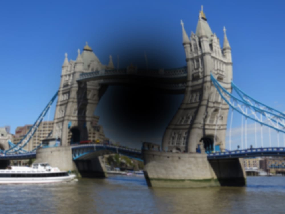
Glaucoma is a chronic eye disease that damages the optic nerve and can result in permanent vision loss. The condition is often caused by elevated pressure inside the eye when fluid drainage becomes blocked. Researchers estimate that glaucoma affects approximately 76 million people worldwide.

One treatment option is the MicroShunt, a tiny filtration device surgically implanted in the eye to help excess fluid drain more effectively. Compared with traditional glaucoma surgeries, the MicroShunt is associated with fewer post-operative complications and often reduces the need for ongoing medication.

Why the Implant Material Can Be Affected

The MicroShunt is manufactured from a styrenic thermoplastic elastomer made from a polystyrene-block-polyisobutylene-block-polystyrene (SIBS) block polymer. This material is designed to be flexible, highly biocompatible, and less likely to cause inflammation or scarring inside the eye.

At the same time, the material is sensitive to contact with hydrocarbon- and oil-based substances. Because it has a strong affinity for oils, petrolatum-based eye ointments can penetrate the implant. When oil components enter the material, the device may swell and experience changes in its shape and mechanical strength.

Manufacturer Warnings Often Overlooked

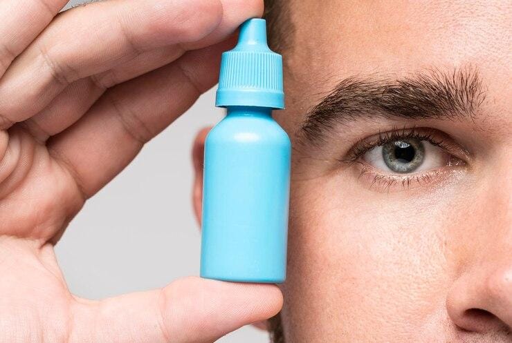
The MicroShunt manufacturer specifically cautions against this type of exposure. According to the instructions, “the MicroShunt should not be subjected to direct contact with petrolatum-based (i.e., petrolatum jelly) materials, such as ointments and dispersions.” Despite this guidance, the warning is not always widely recognized or consistently followed in clinical settings.

“Swollen MicroShunts can be structurally fragile,” said ophthalmologist and Assistant Professor Ryo Tomita of Nagoya University Graduate School of Medicine, the study’s first author. “During surgery, I observed a rupture in a swollen MicroShunt. If more clinicians are aware of this risk, they will be able to prevent similar problems.”

Collaboration Between Medicine and Engineering

To investigate the issue more closely, Tomita worked with Assistant Professor Taiga Inooka and Associate Professor Kenya Yuki from Nagoya University Hospital and the Graduate School of Medicine. They collaborated with Dr. Takato Kajita and Junior Associate Professor Atsushi Noro from the Graduate School of Engineering to study how the MicroShunt changes after contact with petrolatum-based eye ointments.

The medical team analyzed patient cases, while the engineering researchers carried out laboratory experiments. The results were published in Graefe’s Archive for Clinical and Experimental Ophthalmology.

Clinical Evidence From Patient Cases

The clinical analysis involved seven glaucoma patients whose MicroShunt implants were later removed for various reasons. A clear pattern emerged based on whether the implant had been exposed to petrolatum-based ointment.

In three cases, the MicroShunt was exposed outside the conjunctiva and patients were treated with a petrolatum-based eye ointment. All three devices showed noticeable swelling, and two of them had ruptured.

In another three cases, the MicroShunt remained covered by the conjunctiva and no ointment was used. These implants maintained their original structure.

One additional case was particularly revealing. Although the MicroShunt was exposed outside the conjunctiva, no ointment was applied. In this case, the implant did not swell. This finding indicates that direct contact with the ointment, rather than conjunctival exposure alone, is the primary cause of swelling.

Laboratory Tests Confirm the Mechanism

Laboratory experiments reinforced the clinical findings. Researchers immersed unused MicroShunts in petrolatum-based eye ointment to recreate the changes observed in patients.

Microscopic measurements revealed rapid expansion. After 24 hours of exposure, the outer diameter of the MicroShunt increased to 1.44 times its original size. The fin-like portion of the device widened to 1.29 times its initial dimension.

Chemical testing explained why these changes occurred. After 24 hours of immersion, oil components accounted for about 45% of the MicroShunt’s total weight. After three months, oil content increased to 73%.

These results confirmed that swelling is driven by the absorption of oil-based ointment components into the implant material.

Implications for Glaucoma Treatment

Based on their findings, the researchers advise clinicians to avoid using petrolatum-based eye ointments in patients with MicroShunt implants, especially when the device is exposed outside the conjunctiva. They recommend considering alternative post-operative treatments and note that further studies are needed to determine whether swelling affects implant performance even when rupture does not occur.

“Our study found that commonly used medical materials can cause unexpected complications if their chemical properties and usage environments are not fully understood,” Noro stated. “From both medical and engineering perspectives, we emphasize the importance of understanding the chemical properties of medical materials and appropriately managing their usage environments.”


Tips-for-Protecting-Your-Eyes-1200x450.png?strip=all&lossy=1&ssl=1

May month is devoted to raising awareness of eye health issues, which includes preventive measures that help individuals optimize their vision, empowering them to safeguard their eyes and live a happy life. During Healthy Vision Month, we want to share with you some of the most effective tips for protecting your vision. So, let us begin without wasting any time.


Eye-drops-recalled.jpg?strip=all&lossy=1&ssl=1

This year has spooked consumers like no other, with multiple warnings from the FDA about dozens of eye drops that contain potential bacteria and contamination. In January and February, the Food and Drug Administration recalled two over-the-counter eye drops-EzriCare Artificial Tears and Delsam Pharma's Artificial Tears, after they were found to cause bacterial contamination. An outbreak of a rare drug-resistant strain of Pseudomonas aeruginosa was linked to the usage of these eye drops. Out of 55 patients in 12 states who use these drugs, one patient died of a bloodstream infection.


macular-degeneration-1200x675.jpg?strip=all&lossy=1&ssl=1

With age, you might have difficulty focusing on objects up close. You may become more sensitive to glare and have trouble adapting to different levels of light. Let’s look at some of the more common eye problems related to aging:


iokanan_vfx_v21-1200x675.jpg?strip=all&lossy=1&ssl=1

Genetic and clinical research has revealed a new form of macular dystrophy (A disease that causes central vision loss)

Researchers from the National Eye Institute (NEI) have identified a new disease that affects the macula, a small part of the light-sensing retina needed for sharp, central vision. Scientists report their findings on the novel macular dystrophy, which is yet to be named, in JAMA Ophthalmology. NEI is part of the National Institutes of Health.

Macular dystrophies are disorders that usually cause central visual loss because of mutations in several genes, including ABCA4, BEST1, PRPH2, and TIMP3.

For example, patients with Sorsby Fundus Dystrophy, a genetic eye disease specifically linked to TIMP3 variants, usually develop symptoms in adulthood. They often have sudden changes in visual acuity due to choroidal neovascularization- new, abnormal blood vessels that grow under the retina, leaking fluid and affecting vision.

TIMP3 is a protein that helps regulate retinal blood flow and is secreted from the retinal pigment epithelium (RPE), a layer of tissue that nourishes and supports the retina’s light-sensing photoreceptors. All TIMP3 gene mutations reported are in the mature protein after it has been “cut” from RPE cells in a process called cleavage.

“We found it surprising that two patients had TIMP3 variants not in the mature protein, but in the short signal sequence the gene uses to ‘cut’ the protein from the cells. We showed these variants prevent cleavage, causing the protein to be stuck in the cell, likely leading to retinal pigment epithelium toxicity,” said Bin Guan, Ph.D., lead author.

The research team followed these findings with clinical evaluations and genetic testing of family members to verify that the two new TIMP3 variants are connected to this atypical maculopathy.

“Affected individuals had scotomas, or blind spots, and changes in their maculas indicative of disease, but, for now, they have preserved central vision and no choroidal neovascularization, unlike typical Sorsby Fundus Dystrophy,” said Cathy Cukras, M.D., Ph.D., a Lasker tenure-track investigator and medical retina specialist who clinically evaluated the patients.

NEI’s Ophthalmic Genomics Laboratory gathers and manages specimens and diagnostic data from patients who have been recruited into multiple studies within the NEI clinical program to facilitate research of rare eye diseases, including Sorsby Fundus Dystrophy.

“Discovering novel disease mechanisms, even in known genes like TIMP3, may help patients that have been looking for the correct diagnosis, and will hopefully lead to new therapies for them,” said Rob Hufnagel, M.D., Ph.D., senior author and director of the Ophthalmic Genomics Laboratory at NEI.

The study was funded by the NEI Intramural Research Program.


how-eyes-detect-quantity-goodrich-optical.jpg?strip=all&lossy=1&ssl=1

If you can count, it's thanks to your pupils. The holes in the center of your eyes can detect quantity, new research finds. The mechanisms we use to sense quantity are located in our pupils. This is the result of a study conducted by the School of Psychology of the University of Sydney, in collaboration with the Universities of Pisa and Florence (Italy), just published in Nature Communications.


Optimized-freeform-progressive-lenses-goodrich-optical-1200x752.jpg?strip=all&lossy=1&ssl=1

What are the Advantages of Having Optimized Free Form Progressive Lenses in My Glasses? Optimized Free Form progressive lenses from Goodrich Optical are becoming far more popular these days. Primarily because of their visual performance. Couple that with their custom position of wear characteristics and you have unmatched optical performance from your eyewear.


16_Tower-Bridge.jpg?strip=all&lossy=1&ssl=1

New strategy for treating common retinal diseases shows promise A potential treatment based on a natural protein may offer broader benefits than existing drugs Scientists at Scripps Research have uncovered a potential new strategy for treating eye diseases that affect millions of people around the world, often resulting in blindness.


Goodrich Optical Logo


Contact us


Visit us anytime

Goodrich Optical, 2450 Delhi Commerce Dr. Holt, MI 48842


Send us an email

info@goodrichoptical.com


Subscribe


Sign up for our newsletter to receive all the latest eye health news as well as offers and discounts from Goodrich Optical.


    Copyright © 2023 all rights reserved.


    Copyright ©2022 all rights reserved